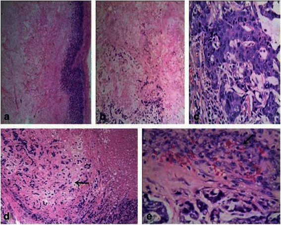
Figure 3.

Microscopical findings of CNC. (a): The tumor presented with an extensive central necrotic or acellular zone surrounded by ring-like or ribbon-like residual tumor tissue. The transition between the central necrotic or acellular zone and the viable tumor tissue was abrupt (×4). (b): The central zone of the tumor showed coagulative necrosis with pink, fine granules combined with fibrotic or hyaline material and a tumorous stroma around the central necrotic zone, accompanied by myxoid matrix formation (×10). (c): The tumor cells were arranged in cord-like or nest-like patterns. Most of the tumor cells showed evident atypia, prominent nucleoli, and frequent mitotic figures (×40). (d): Focal cartilaginous metaplasia was present in this case (→)(×10). (e): The periphery of the tumors demonstrating the granulomatous reaction(→) (×40).
