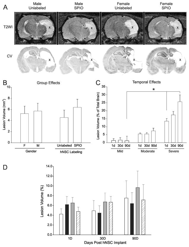
Figure 3. Lesion volumes are not influenced by gender or hNSC labeling.
A) At 90d post HII T2WI lesions (hyperintensities, X) appeared similar across all gender and implant groups. Cresyl violet post-mortem histology (lower panel) showed similar lesion size to T2WI (upper panel). Cystic lesions (X) were similar between gender and hNSC labeled animals; B) Lesion volumes are not altered by gender or labeling; C) Lesion volume evolution over time did not change in mildly injured animals, but there was an increase in lesion volume in moderate and severe HII injury groups (see text). Across all groups there was a significant difference between severity groups at 1 and 90d (p=0.05). D) No significant differences were found in lesion volumes over the 90d experimental period by gender or labeling. (Comparison of gender, labeling status and lesion volume, p=0.684) (white bar, M Unlabeled, black bar, F Unlabeled, gray bar, M SPIO; hatched bar, F SPIO)

