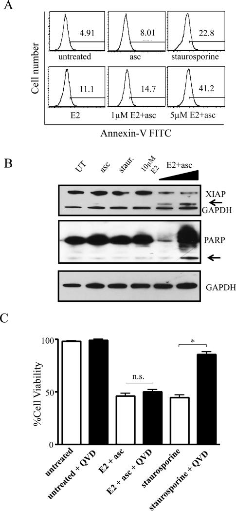
Figure 5. Cell death caused by MnP+asc shows classical markers of apoptosis, but is caspase-independent.
A) SUM149 cells were treated with sublethal doses (corresponding to EC10 and EC25, respectively) of E2 ± ascorbate or ascorbate alone for 4h and stained with Annexin-FITC. Histograms shown are representative of 2-3 independent experiments. Staurosporine was used as a control. B) Western immunoblot analysis of X-linked inhibitor of apoptosis (XIAP, top panel) and poly-ADP ribose polymerase (PARP, middle panel) in SUM149 cells treated with ascorbate or E2, alone and in combination. E2 doses in combination with 3.3 mM ascorbate: 1 μM and 10 μM. GAPDH was used as loading control. Arrows denote cleavage products. C) Viability as determined by trypan blue exclusion assay of SUM149 cells treated with 10 μM E2+ascorbate or staurosporine for 24 hours, alone (white bars) or after 30 minute pre-treatment with 20 μM QVD-OPh (black bars), a pan-caspase inhibitor. Data represent mean±SEM viable cells taken as a percentage of total cells (n=2-3, *p<0.05).

