Figure 2.

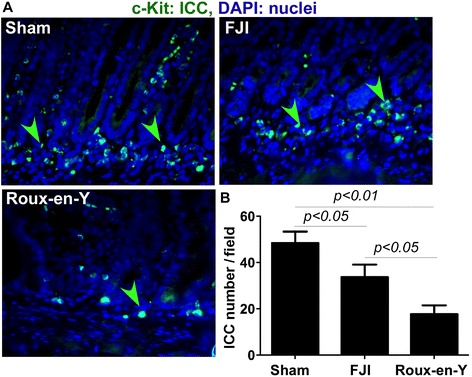
FJI preserves ICC in the small intestine. The intestinal tissues were isolated from beagles at 48 hours postoperatively. Paraffin-embedded tissue sections were stained using an anti-c-Kit (ICC marker) antibody and a FITC-conjugated secondary antibody. Nuclei were stained using DAPI (blue). Fluorescent and DAPI images were taken from the same field (A). Green arrowheads: ICC. The number of ICC per 20X power field is shown (B). Sham = 5 beagles, FJI = 10 beagles, Roux-en-Y = 7 beagles.
