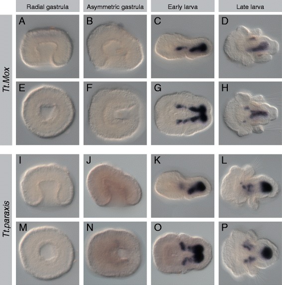
Figure 7.

Expression patterns of Tt.Mox and Tt.paraxis. All images are oriented with anterior to the left. Panels (A-D) and (I-L) are lateral views. Panels (E, F) and (M, N) are blastoporal views. Panels (G, H) and (O, P) are ventral views. For detailed descriptions of expression patterns, see text. (A-H) Tt.Mox expression is first detected in the early larva in lateral mesodermal bands flanking the endoderm. Expression in the late larva is in the ventromedial mesoderm. (I-P) Expression of Tt.paraxis is first detected in the early larva, in the mesoderm of the mantle and pedicle lobes. In the late larva, there are distinct domains of expression underlying the mantle lobe and in the posterior mesoderm of the pedicle lobe.
