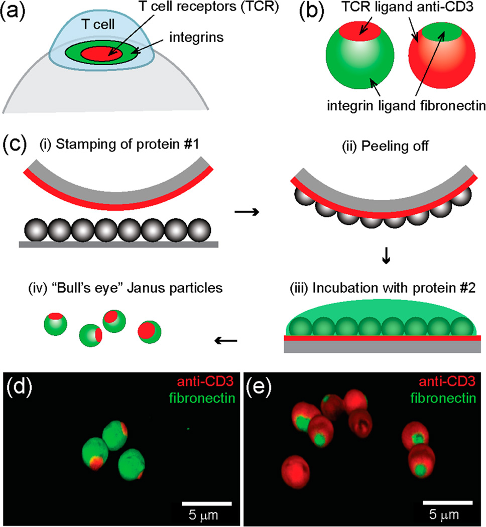
Figure 1.
“Bull’s eye” Janus particles resemble the spatial pattern of proteins found in the immunological synapse. (a) Ligand-bound T cell receptors (TCRs) and integrins form “bull’s eye” concentric microdomains in the membrane junction between a T cell and an antigen-presenting cell, which is known as the immunological synapse. (b) Schematic illustration of native and reverse “bull’s eye” particles that display patterns of anti-CD3 and fibronectin. (c) Microcontact printing method for creating “bull’s eye” particles. (d, e) 3-D confocal fluorescence images of the “bull’s eye” particles.

