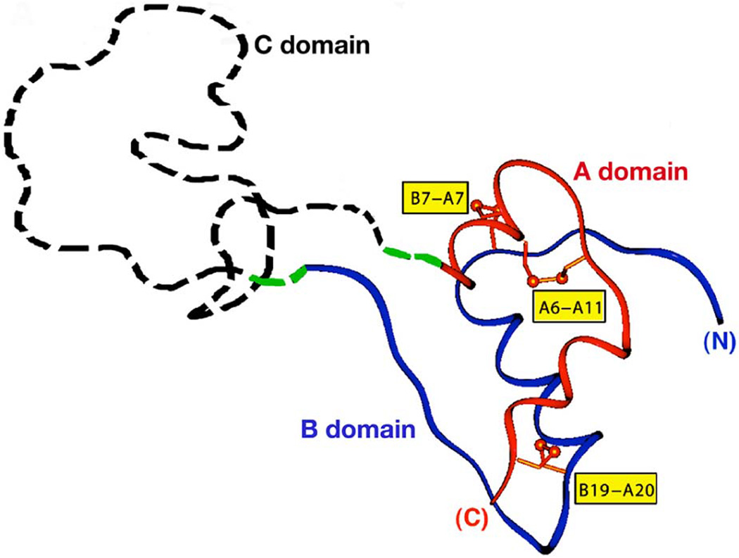
Figure 1. Structure of proinsulin.
Proinsulin is comprised sequentially of insulin B domain (blue), connecting C domain (which is disordered and therefore has no fixed structure), and insulin A domain (red). Six cysteines form three disulfide bonds that are labeled in yellow boxes. This figure was adapted from Liu, M., et al. 2010. PLoS ONE. 5(10):e13333.

