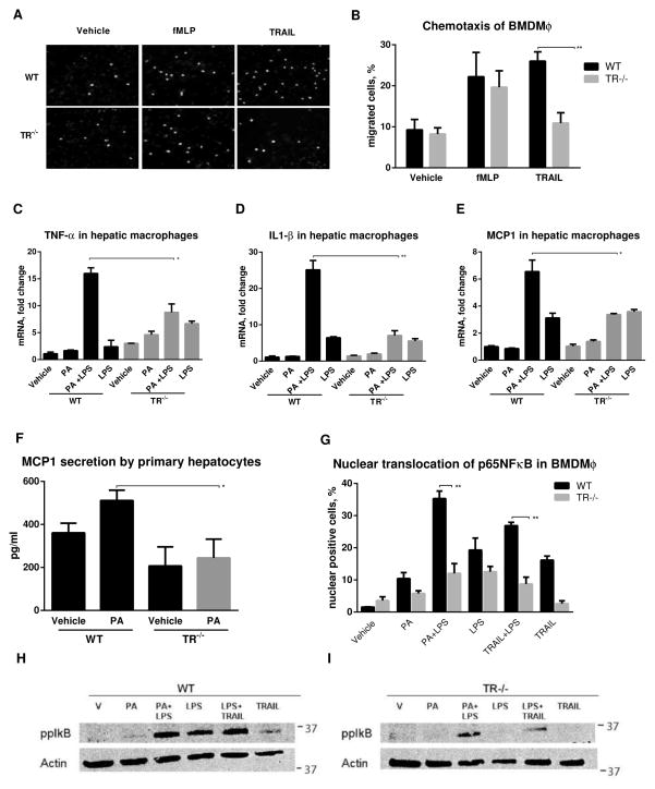
Fig. 4. Macrophage TR signaling contributes to the inflammation of nutrient excess.
(A) Fluorescent photomicrographs (scale bar 20 μm) and (B) quantification of DAPI stained migrated WT and TR−/− BMDMϕ. (C–E) mRNA abundance of Tnfα, Il1β, and Mcp1 in cells treated with 400 μM palmitate (PA) and/or 10 ng/ml lipopolysaccharide (LPS), 8 h. (F) MCP1 levels in supernatants from cells treated with 400 μM PA, 8 h. (G) Quantification of nuclear translocation of NF-κB by immunofluorescence, (H) phosphorylation of IκB-α (Ser32/Ser36) in WT and TR−/− BMDMϕ treated with 400 μM PA ± 10 ng/ml lipopolysaccharide (LPS) or 10 ng/ml TRAIL for 1 h. *p<0.05, **p<0.01

