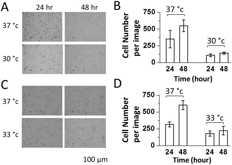
Fig 1. Bright-field images (34.8x magnification) of HEK-293S cells growing at 37°C (upper A and C), 30°C (lower A), and 33°C (lower C).
HEK-293S cells were seeded at a density of 5.5 x 10⁵ cells/35 mm Petri dish. The images were taken at 24 and 48 hours using a digital camera mounted on a Carl Zeiss Axiovert S200 microscope (see Methods). Cell number counts per image or 0.22 mm2 were plotted in (B) and (D) based on the images shown in (A) and (C), respectively. For each dish, 3 viewing areas were randomly chosen and their images were taken.

