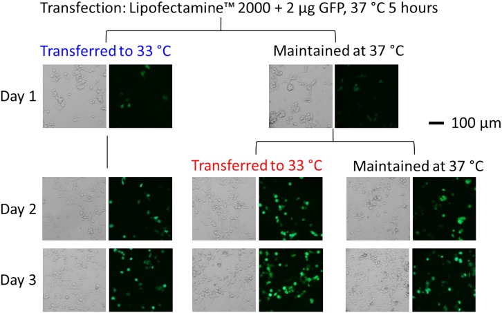
Fig 2. Bright-field and fluorescence images (34.8x magnification) of GFP-expressing HEK-293S cells.
The cells were transfected with the GFP plasmid using Lipofectamine 2000. Five hours after transfection (the cells were maintained in a 37°C incubator during this 5-hour period), the medium was replaced. Three sets of dishes were subject to three different ways of maintaining culture temperature, as shown. One set of dishes was transferred to a 33°C incubator. Another set was returned to the 37°C incubator; 19 hours later or 24 hours after transfection, this set of dishes was brought to the 33°C incubator and remained there. The control dishes were maintained at 37°C throughout the experiment. The images were taken from the first day (24 hours after transfection) to the third day.

