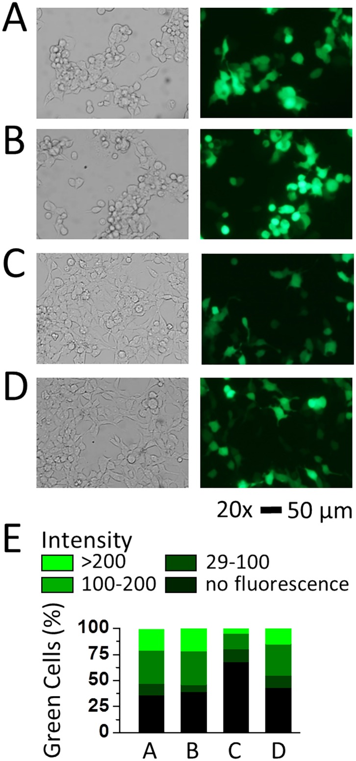
Fig 4. Transfection of the GFP plasmid in HEK-293S cells with different reagents and the expression of GFP at 33°C.
Bright-field and fluorescence images (34.8x magnification, scale bar = 50 μm) of HEK-293S cells taken at the 48th hour after transfection. Transfection of HEK-293S cells as in images (A) to (D) was carried out using Lipofectamine 2000, Lipofectamine LTX & PLUS, Metafectene EASY with Opti-MEM and Metafectene EASY with DMEM with 10% FBS, respectively. In each of the transfections, 2 μg GFP plasmid for a 35 mm Petri dish was used. On day 1, all transfected dishes were incubated at 37°C; on day 2, these dishes were transferred to a 33°C incubator. Based on these images, the transfection efficiency for transient expression of GFP was determined to be ~64%, ~61%, ~32% and ~57% from (A) to (D), respectively. (E) The green cells were categorized into high (intensity > 200), middle (intensity 100–200), and low (intensity 29–100) three groups. The black color indicates cells visible in the bright view but can’t been observed under UV (intensity < 29). The percentage of each group was plotted in stacked columns.

