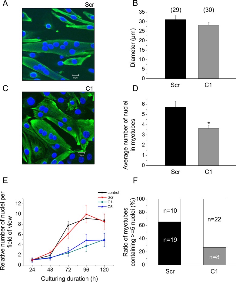
Fig 7. Effect of decreased SERCA1b expression on proliferation and differentiation of C2C12 cells.
Immunocytochemical staining of terminally differentiated C2C12 myotubes demonstrating the morphological changes (C) in cloneC1 compared to (A) scrambled shRNA transfected cells. Muscle specific desmin was detected and visualized with FITC-conjugated secondary antibody. Nuclei were stained with DAPI. Images were recorded from1 μm thick optical slices. Original magnification was 40×. (E) Proliferation rate was calculated from the increase in the number of myogenic nuclei after normalising to the value obtained after 24 hours of culturing. Data represent mean ± standard error of the mean (SEM). (B,D, and F) Quantitative parameters of differentiated multinucleated myotubes.CloneC1 and scrambled shRNA transfected cells were compared. Numbers in parentheses indicate the number of identified myotubes on 3 different coverslips. Asterisks (*) mark significant (P<0.01) differences.

