Figure 4.

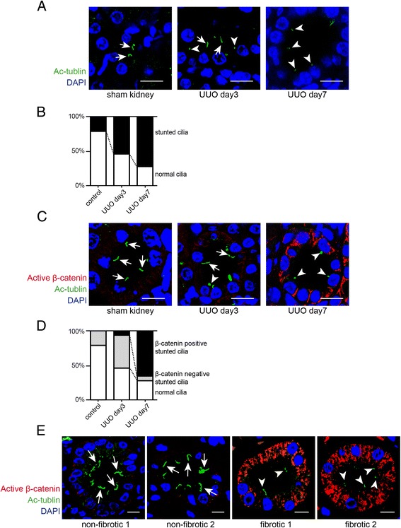
Stunted cilia and canonical Wnt signaling in experimental obstructive nephropathy. (A) Paraffin-embedded kidney sections were labeled with antibodies to acetylated α-tubulin (Ac-α-tubulin) (green). Nuclei were labeled with DAPI (blue). Staining was analyzed using a confocal microscope. The pictures display representative confocal photomicrographs of labeled sections of sham-operated mice (left) and of kidneys 3 days (middle) and 7 days (right) after ureter obstruction. Scale bars: 10 μm. Arrows indicate normal cilia and arrowheads indicate stunted cilia. (B) The graph summarizes the relative distribution of normal and stunted cilia in each group. (C) Immunofluorescence double-labeling experiments were performed using specific antibodies to acetylated α-tubulin (cilia) and non-phosphorylated β-catenin (active canonical Wnt signaling). Nuclei were stained with DAPI. The pictures display representative photomicrographs of sections from sham-operated control mice (left) and post-obstructive (days 3 and 7) kidneys. Non-phosphorylated β-catenin immunostaining was most abundant in tubules with stunted cilia. Arrows indicate normal cilia and arrowheads display stunted cilia. Scale bars: 10 μm. (D) The graph shows relative distribution of normal cilia and stunted cilia regarding to β-catenin expression in each group. (E) Non-fibrotic and fibrotic kidney sections from human biopsies were labeled with acetylated α-tubulin (cilia) and non-phosphorylated β-catenin. Pictures display representative confocal photomicrographs of each sample. Arrows indicate normal cilia and arrowheads indicate stunted cilia. Scale bars: 10 μm. UUO, unilateral ureteral obstruction; DAPI, 4′,6-diamidino-2-phenylindole.
