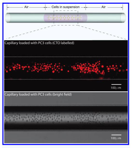
Figure 2.

A schematic drawing of the loaded cells in capillary (Top). PC3 labeled with Cell Tracker Orange (CTO) epifluorescence in Red (Middle). Bright field image of the cells in capillary (Bottom).

A schematic drawing of the loaded cells in capillary (Top). PC3 labeled with Cell Tracker Orange (CTO) epifluorescence in Red (Middle). Bright field image of the cells in capillary (Bottom).