Fig. 6.
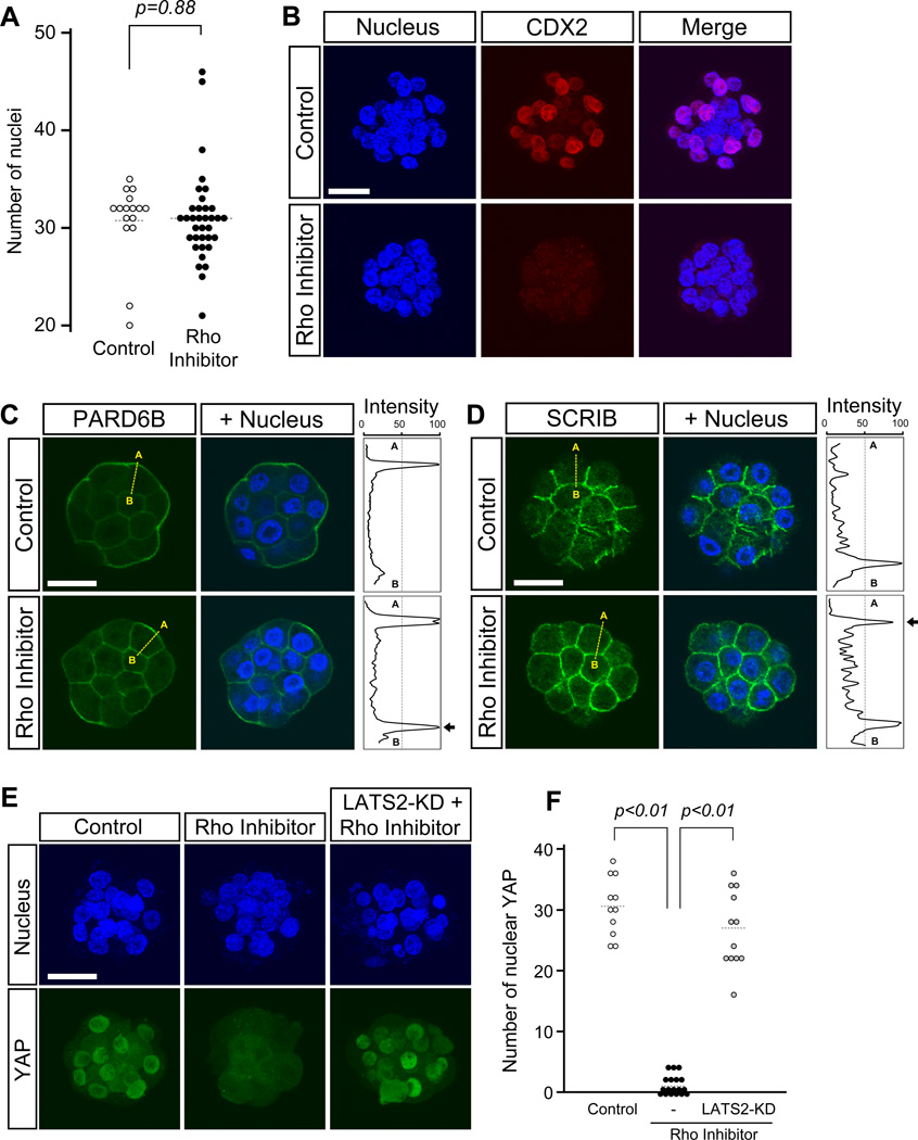
RHO inhibition phenocopies the Y-27632-induced disruption of cell polarity and activation of Hippo signaling in embryos. Embryos were cultured in the absence or presence of RHO Inhibitor I from 8- to 32-cell stage (E2.5–E3.5). (A) Comparison of total nuclear numbers. There is no statistically significant difference (Student t-test) between control (n=16) and inhibitor-treated (n=35) embryos. Circles represent the number of DAPI-stained nuclei in individual embryos, and horizontal dashed bars represent the mean value for each group. (B) Immunostaining for the TE lineage marker, CDX2 (red), in representative embryos. CDX2 is diminished in the inhibitor-treated embryo. Images are Z-axis projections of optical sections. (C-D) Immunostaining for apical-basal polarity proteins in representative embryos. In the control embryo, PARD6B (green in C) is strongly enriched in the apical domains and weakly enriched in the basolateral domains of outer cells. With inhibitor treatment, PARD6B localization becomes more distinct in the basolateral region. On the other hand, SCRIB (green in D) is enriched in the basolateral domains of outer cells in the control embryo, but also becomes localized in the apical domains in the treated embryo. Images are optical sections. Abbreviations: A (apical), B (basal). (E) Nuclear YAP (green) diminishes in the inhibitor-treated embryo. Overexpression of dominant-negative LATS (LATS2-KD) rescues nuclear localization of YAP in the inhibitor-treated embryo. Images are Z-axis projections of optical sections. (F) Comparison of the number of YAP-positive nuclei. Nuclear localization of YAP is significantly reduced in RHO inhibitor-treated embryos (n=18) compared to untreated control embryos (n=11; Student t-test). Nuclear localization of YAP is restored in inhibitor-treated embryos that have been overexpressed with LATS-KD (n=12; Student t-test). Circles represent the number of YAP-positive nuclei in individual embryos, and horizontal dashed bars represent the mean value for each group. Blue, DAPI. Scale bars: 20 µm.

