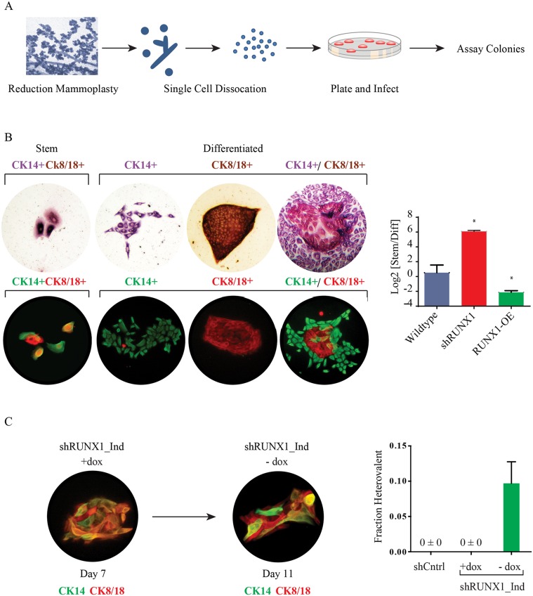
Fig 8. RUNX1 inhibition blocks the differentiation of patient-derived mammary stem cells.
(A) Schematic of the human breast progenitor cell colony formation assay. Human primary organoids were dissociated into single cells and plated onto culture plates. (B) Brightfield and fluorescent images of CK14 and CK8/18 stained human progenitor cell colonies: stem cell colonies are small and consist of cells dual positive for CK8/18 (brown;red) and CK14 (red/purple; green); differentiated colonies are larger and have domains that are singly positive for CK8/18 and CK14. Quantification of progenitor cell colonies from control, shRUNX1 or RUNX1 overexpressing cells; (C) Immunofluorescence and quantification of heterovalent colonies formed by primary human cells infected with a dox-inducible shRUNX1 in the presence of dox for 7 days. After 7 days, dox was removed and the colonies were grown for an additional 96 hours. These cultures generated heterovalent colonies—defined as colonies containing both stem and differentiated cell types. Shown is a heterovalent colony containing both CK14,CK8/18 double-positive cells, and cells that only stained for either CK14 or CK8/18. The fraction of heterovalent colonies in each condition is shown. * indicates p<0.05 versus wild type; error bars are SEM with n = 4.

