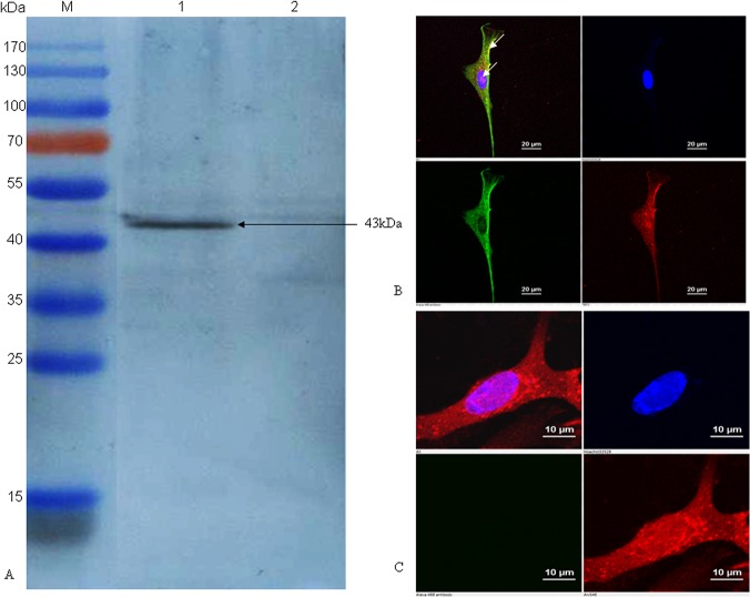
Fig 1. Confirmation of PmpD-N protein expression in HVT vector by immunoblotting assay and indirect immunofluorescence.
(A) The PmpD-N expression in rHVT-pmpD-N was detected by immunoblot with C. psittaci strain 6BC-specific polyclonal antibodies. Lane M, pre-stained protein ladder; Lane 1, cell lysate post inoculation with rHVT-pmpD-N; Lane 2, cell lysate post inoculation with parental HVT. The black arrow indicates the approximately size of 43kDa. (B) Indirect immunofluorescence analysis of PmpD-N expression in CEF cells. CEF cells on glass coverslips were infected with rHVT-pmpD-N, then incubated with mouse anti-PmpD-N polyclonal antibody of C. psittaci and chicken anti-HVT polyclonal serum, and then reacted with goat anti-mouse IgG conjugated with Alexa Fluor 488 (green fluorescence, shown in the lower left panel) and goat anti-chicken IgY labelled with Alexa Fluor 568 (red fluorescence, shown in the lower right panel), respectively. Finally, cell nuclei were stained with DAPI (blue fluorescence, shown in the top right panel). The merged image is shown in the top left panel. The expression of the targeted protein is indicated by white arrows in top left panel. (C) Parental HVT control. CEF cells on glass coverslips were infected with parental HVT, and then the process of test and the panel meaning are the same as those shown in Fig 1B.

