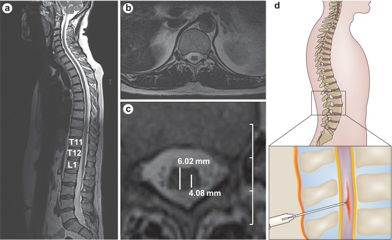
Fig. 1.

Accurate anatomical targeting of stem cell delivery. a. T2-weighted magnetic resonance imaging scan showing a sagittal view of the spinal cord and the position of the conus medullaris and lumbar enlargement. b. Axial view of the spinal cord at the level of T12. c. Precise needle placement into the ventral horn of the spinal cord is calculated from a magnified image of part b. Estimated measurements of spinal cord diameter (6.02 mm) and distance from the dorsal root entry zone to the ventral horn (4.08 mm) are shown. Scale: 1 cm per grid division. d. Schematic of targeted injection of stem cells into the spinal cord. Reproduced from Boulis et al., Nat Rev Neurol 2011;8:172–6, [10]
