Fig. 1.
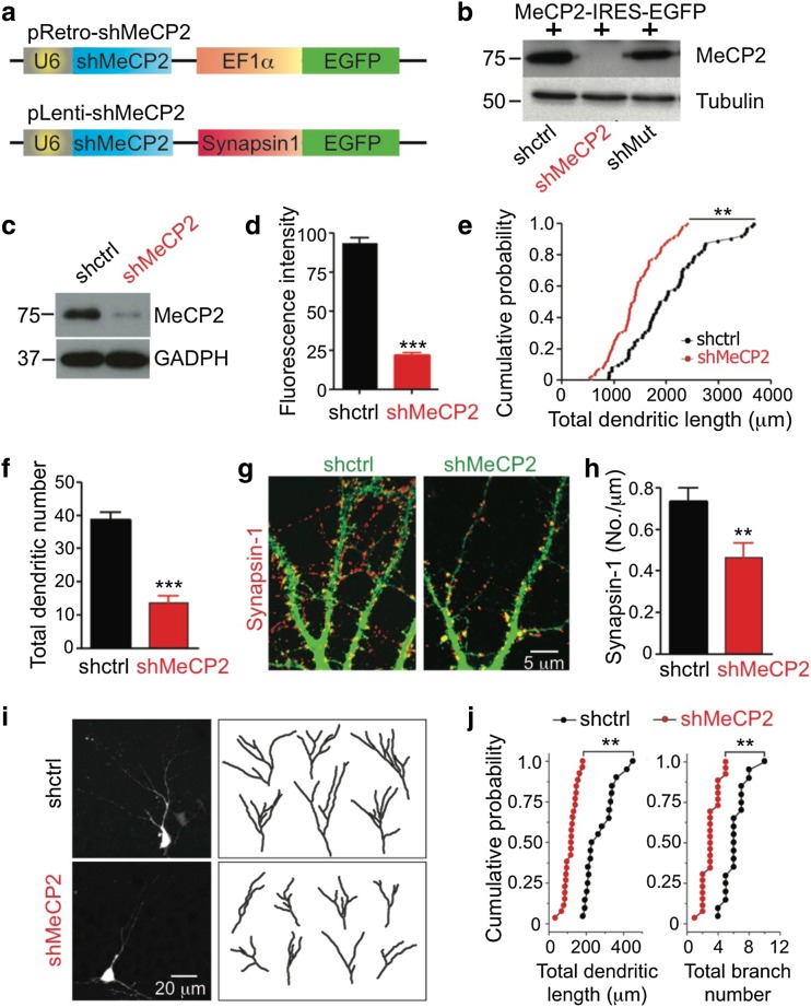
Methyl-CpG-binding protein 2 (MeCP2) deficiency affects dendritic development and synaptogenesis in hippocampal neurons. (a) Schematic diagram showing the knockdown retroviral and lentiviral constructs. EF1α = elongation factor 1- α; EGFP = enhanced green fluorescent protein. (b) Representative Western blot showing the efficiency of retrovirus carrying short hairpin RNA (shRNA) of scrambled sequence or shRNAs against MeCP2 (shMeCP2, shMut) in knocking down MeCP2 in human embryonic kidney 293 cells overexpressing MeCP2 using Western blot analysis at 5 days in vitro (DIV). IRES = internal ribosomal entry site. (c) Representative Western blot showing the efficiency of lentivirus with shMeCP2 in knocking down MeCP2 in embryonic day 18 (E18) primary hippocampal neurons at 14 DIV. GAPDH = glyceraldehyde 3-phosphate dehydrogenase. (d) Average fluorescence intensity per E18 hippocampal neurons (at 5 DIV) infected with lentivirus carrying scrambled RNA (shctrl) or shMeCP2 (***p < 0.01; Student’s t test) (e) Quantification of total dendritic length and (f) total dendritic branch number of shctrl- or shMeCP2-expressing cells (***p < 0.01; Student’s t test). (g) Representative images of neurons expressing shctrl or shMeCP2 (green), immunostained with anti-synapsin I (red) (scale bar = 5 μm). (h) Density of synapsin-1 positive puncta per μm dendrite in hippocampal neurons transfected with shctrl or shMeCP2 (**p < 0.01; Student’s t test). (i) Representative images and tracings of dendritic arbors of dentate gyrus granule neurons infected with retrovirus carrying shctrl (upper panel) or shMeCP2 (lower panel) in vivo (scale bar = 5 μm). (j) Quantitative analysis of the (left) total dendritic length, (right) total branch number of control, and MeCP2 deficient neurons (**p < 0.01; Student’s t test)

