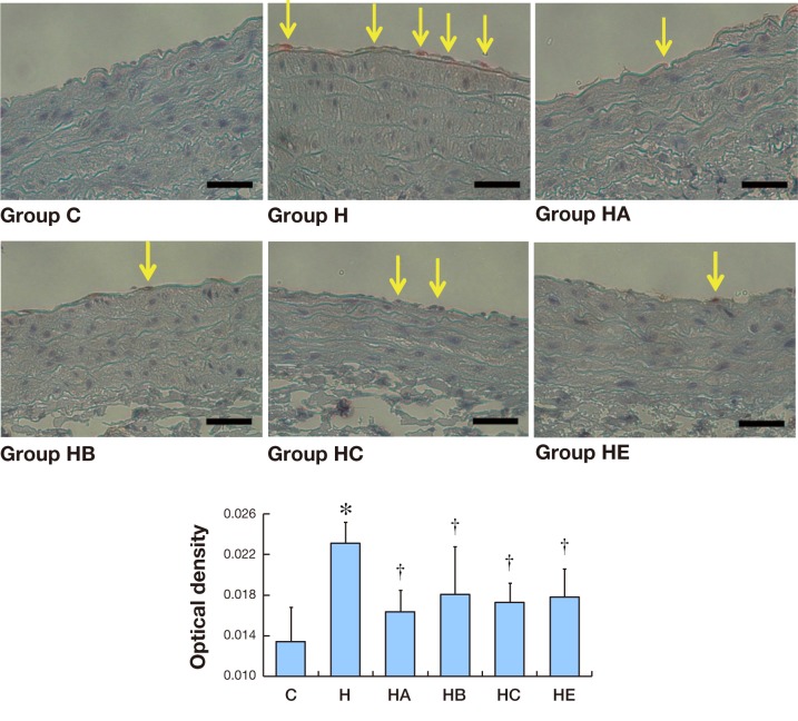
Fig. 5.
Immunohistochemistry expression of ICAM-1 in aorta.
A: Seldom ICAM-1 immunostaining (red-brown deposits indicate positive staining) is found on the whole layers of abdominal aortas in Group C. Bars express 25 μm.
B: Mean optical density values of ICAM-1. The photographs generated were quantitatively analyzed the optical density of ICAM-1 with Image-Pro Plus version 6.0 software. *P < 0.05 versus Group C. †P < 0.05 versus Group H.

