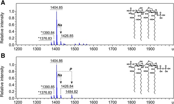
Figure 2.

Charge deconvoluted ESI FT-ICR mass spectra in negative ion mode of lipid IVA isolated from BW30270-derived mutants. The lipid IVA (calculated mass 1404.854 u) was extracted from E. coli (K-12) strains KPM318 (A) and KPM335 (B). Mass numbers given refer to the monoisotopic masses of neutral molecules. Peaks representing molecules with variations in acyl chain length are labeled with asterisks (∆m = 14.02 u). The molecular ion at 1484.82 u in panel B indicates the presence of a minor fraction of 1-diphosphate lipid IVA. The structures of lipid IVA and 1-diphosphate lipid IVA are shown as insets in panels A and B, respectively.
