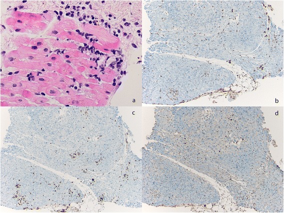
Figure 3.

Histological analysis of endomyocardial biopsy. (a) Hematoxylin and eosin staining of the myocardial biopsy with focal mononuclear infiltrates. (b) Immunohistochemical analysis of CD68 macrophages. (c) Staining for CD8 positive T cells (d) and FOXP3 positive cells within the myocardium of the patient.
