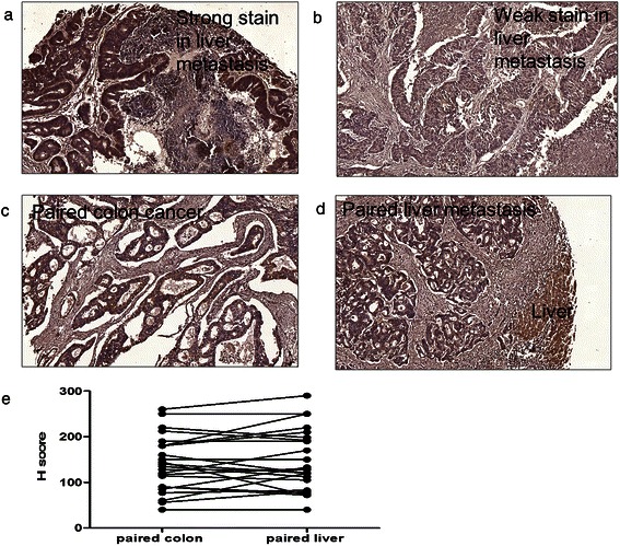
Figure 2.

Immunohistochemical (IHC analysis of CIP2A expression in patients with colorectal cancer. Representative examples of CIP2A expression: (a) strong expression in colorectal liver metastasis, (b) weak expression in colorectal liver metastasis, (c) staining in paired colon cancer, (d) colorectal metastasis tissues, and (e) H-score in paired colon cancer and liver metastasis samples. CIP2A was not consistently overexpressed in colon cancer compared with colorectal liver metastasis in paired tissue specimens.
