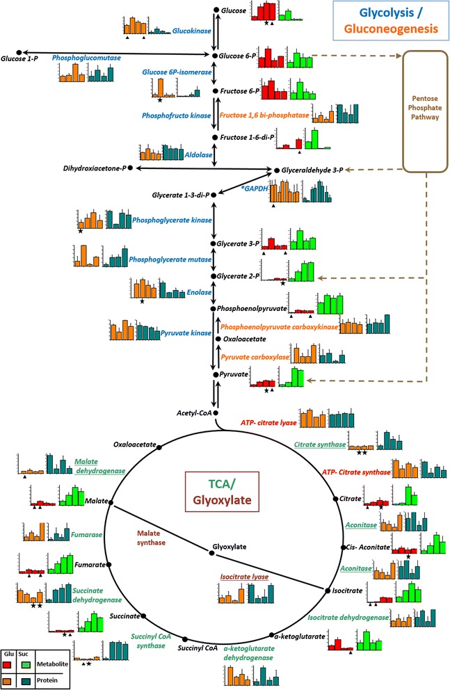
Figure 3.

Protein and metabolite levels in the glycolysis, gluconeogenesis, glyoxylate and TCA pathways. According to the carbon source the mean value and SD of abundance for each protein, and the response ratio for each metabolite were normalized to a value of 100. Each column graphic represents the growth phases studied (from left to the right: lag, early exponential, late exponential and stationary). In the column charts a color code was included to differentiate proteins and metabolites found when X. dendrorhous was cultured with the different carbon sources: proteins in glucose (orange), proteins in succinate (cyan), metabolites in glucose (red) and metabolites in succinate (green). The pathways were adapted from the KEGG database. In the metabolic pathways, names are written in a color letters: metabolites (black), glycolysis proteins (blue), gluconeogenesis proteins (orange), glyoxylate proteins (gray) and TCA proteins (green). Proteins involved in both, the glyoxylate and TCA cycle, are underlined and cytosolic proteins related to acetyl-CoA formation are indicated in red. An asterisk beside the protein name and in the multiple column charts indicates that the protein probably suffers post-translational modifications as it was identified in multiple spots. Statistical significant differences between samples from different carbon sources at the same growth phase are represented as stars (t-test p < 0.02) and triangles (Benjamini-Hochberg correction p < 0.05). Abbreviations: P: phosphate, TCA: tricarboxylic acid cycle.
