Figure 4.

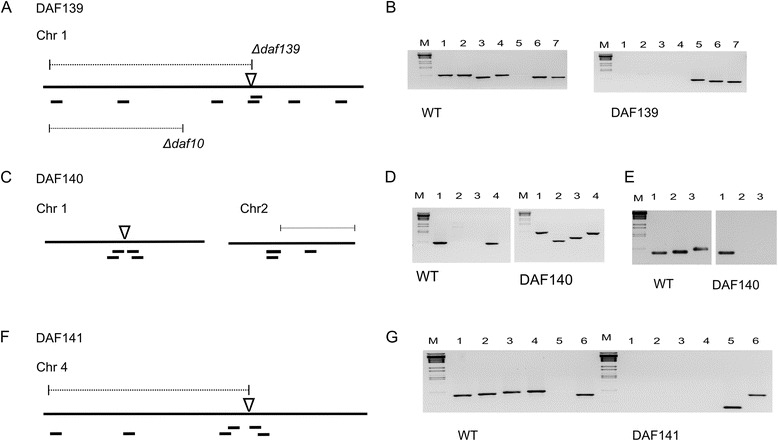
Schematic overview of chromosomal recombination and deletion events for the three DAF mutants and verification by diagnostic PCR. (A) DAF139 partial chromosomal deletion and insertion event. For comparison the chromosomal deletion for mutant DAF10 published earlier by Baldwin et al. [15] is indicated. (B) PCR analysis of chromosomal regions predicted to have either been lost or retained on chromosome 1 in strain DAF139. The wild type and DAF139 genomic DNA was amplified with oligonucleotide pairs in lanes: (1) U516/U517, (2) U518/519, (3) U550/U551, (4) U549/U521, (5) U606/U521, (6) U520/U51 and (7) U522/U523. The approximate location of the amplified PCR fragments is indicated in the schematic overview from left to right corresponding to PCR products in lane 1 to 7. (C) DAF140 shows a single-copy insertion in chromosome 1 and a large untagged deletion on chromosome 2. (D) Diagnostic PCR in lanes: (1) U533/U534, (2) U553/U602, (3) U603/U554, (4) U535/U536. Approximate location of fragments is indicated in the schematic from left (1) to right (4). (E) Diagnostic PCR of wild type and DAF140 chromosome 2 in lanes: (1) U540/U541, (2) U541/U502 and (3) U556/U557. Approximate location of fragments is indicated in the DAF140 schematic from left (1) to right (3). (F) In DAF141 three multimerised plasmids are inserted at position 182,476 bp in chromosome 4. (G) Diagnostic PCR of wild type and DAF141 chromosome 4 in lanes: (1) U600/U601, (2) U558/U559, (3) U545/U552, (4) U545/U546, (5) U604/U605 and (6) U547/U548. Approximate location of fragments is indicated in the schematic from left (1) to right (6). Large black lines indicate the chromosomes (not drawn to scale). Chromosomal deletions (dotted lines), plasmid insertion point (inverted open triangle). Diagnostic PCR fragments (small black lines). DNA ladder BstEII (M).
