Figure 1.

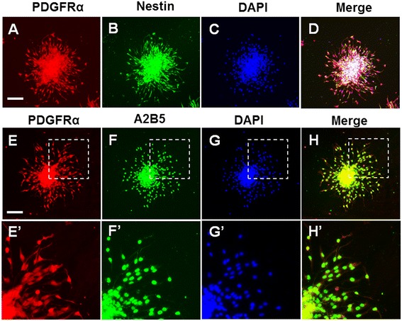
Differentiation of NSC-OPCs. The cells that migrated out of the oligospheres were labeled with antibodies and DAPI. (A) Cell staining with anti-PDGFRα antibody (red); (B) Cell staining with anti-nestin antibody (green); (C) Nuclei staining with DAPI, (D) Merged image of images (A)–(C), (E) Cell staining with anti-nestin antibody (green), (F) Cell staining with anti-A2B5 antibody (green), (G) Nuclei staining with DAPI, (H) Merged image of images (E)–(G). (E’–H’) Magnified images of inset indicated in (E–H), respectively. Scale bar: 100μm.
