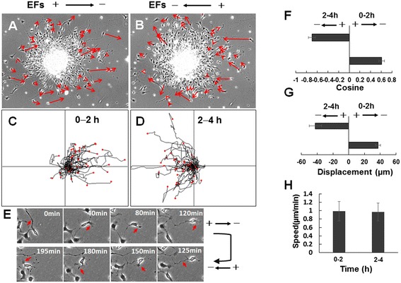
Figure 3.

Reversal of migration direction of ARPC2 +/+ neural stem cell-derived oligodendrocyte precursor cells with reversal of electric field (EF) vectors. (A) Cell migration to cathode pole from oligosphere in EF of 200 mV/mm. (B) Reversed migration of same cells in EF of 200 mV/mm. Arrows show migration direction and positions of cells at beginning and end of each experiment. Scale bar: 100 μm. (C) Tracks of cathodal migration of cells in (A) in EFs. (D) Migration tracks of cells in (B) after EF is reversed. Cells switched direction to migrate to new cathode. (E) Migration of representative cell in EFs. The leading process of the cell guides cell migration toward the cathode during the first 2 hours. Cell migration in opposite direction when EF pole is reversed. (F) Reversal of directedness and (G) reversal of net displacement of cell migration when EF pole is switched to opposite direction. (H) No significant change in cell migration rates before and after EF pole reversal. See Additional file 1: Video 1.
