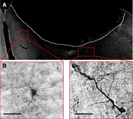
Figure 1.

Two distinct DCX+ cells can be distinguished in the PZ. Representative images of 40 μm brain sections of C57 mice following immunohistochemical staining of DCX. (A), Overview of the circumscribed cortical ischemic infarct. (B), Example of DCX+ cell with multiple processes and weaker immunoreactivity, termed DCX+ stellate cells. (C) The second DCX+ cell type revealed a polar morphology with one or two processes, note the higher signal intensity of DCX. Scale bars 20 μm.
