Figure 1.

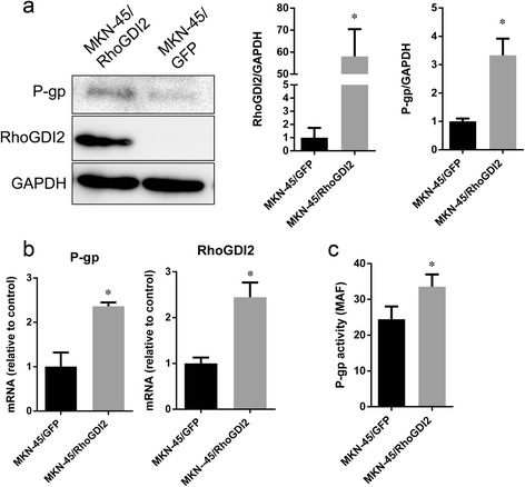
Ectopic expression of RhoGDI2 increased P-gp mRNA, protein and activity. (a) Western blotting analysis of RhoGDI2 and P-gp expression in MKN-45/RhoGDI2 and MKN-45/GFP. The immunoblot image was quantitated by densitometric analysis (right) (n = 3 independent experiments). *p < 0.05 vs MKN-45/GFP. (b) The mRNA of RhoGDI2 (left) and P-gp (right) in MKN-45/RhoGDI2 and MKN-45/GFP was detected by RT-PCR. (c) P-gp activity in MKN-45/RhoGDI2 and MKN-45/GFP was measured as described in materials and methods, MAF was plotted. Data are expressed as mean ± SD from three independent experiments; *p < 0.001 vs MKN-45/GFP.
