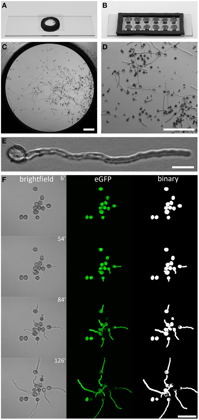
Figure 1.
Arabidopsis pollen imaging in the micro-germination setup. Mounting pollen in a single (A), or multi well (B), micro-germination setup for live cell imaging. In the center of one well (C) PTs grow in close proximity to the cover slip (D), allowing the use of high NA immersion objectives with low working-distances. Pollen germination is not affected by the mounting technique and PTs showed normal morphology (E). A time series of germinating PLat52:GFP pollen is shown in (F). Brightfield images are shown as sum slice projections and confocal fluorescence images of cytoplasmic GFP as maximum intensity projections. Binary images of GFP channel enable unbiased quantitative measurement of PT area size and morphology (F). Scale bars: (C,D) 200 μm, (E) 20 μm, (F) 50 μm.

