Figure 3.
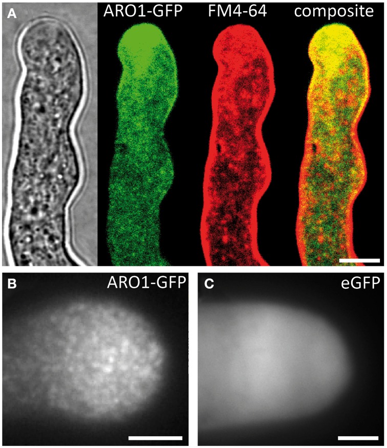
ARO1-GFP localizes to vesicles at the pollen tube tip, accumulating in the inverted cone-shaped region. (A) At the PT tip, ARO1-GFP predominantly accumulates in the vesicle-rich inverted cone-shaped region and partially co-localizes with FM4-64. No co-localization of ARO1-GFP and FM4-64-stained membrane compartments is detected in the subapical region of the PT. (B) TIRF microscopy reveals that ARO1-GFP signals appear as discrete punctate structures of approximately 0.2 μm in the PT tip. These punctate structures are not observed in PTs that express cytoplasmic GFP (C). Scale bars: 5 μm.

