Figure 6.

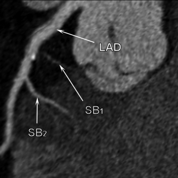
Multi-slice computed tomography examination. Calcifiations are visible on the septal side of the LAD between the origin of first and the second septal branch.

Multi-slice computed tomography examination. Calcifiations are visible on the septal side of the LAD between the origin of first and the second septal branch.