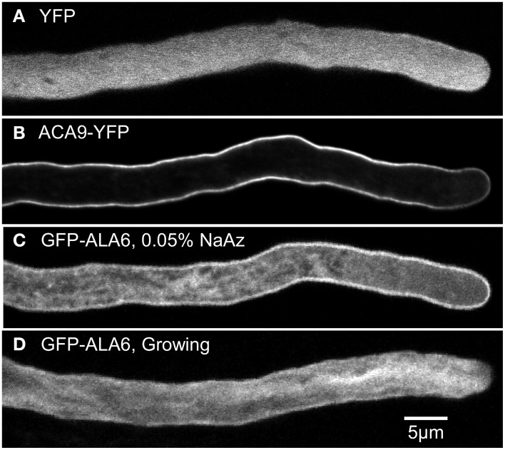
Figure 5.
Confocal fluorescence micrographs showing GFP-ALA6 localizes to the pollen tube perimeter and endomembrane structures. (A,B) Growing pollen tubes expressing (A) YFP (ss1919) as a marker for the cytosol and (B) ACA9-YFP (ss471-472) as a marker for the plasma membrane (Myers et al., 2009). (C,D) Pollen tubes expressing GFP-ALA6 (ss1880) either (C) treated with 0.05% NaAz, or (D) growing. Constructs were expressed under the control of the ACA9 promoter in stable transgenic Arabidopsis plants. Images of GFP-ALA6 are representative of seven GFP-ALA6 (ss1878–1884) and four ALA6-YFP (ss1885–1888) transgenic lines in which the transgenes were shown to rescue the ala6-1/7-2 phenotype. The pattern of localization was equivalent for expression levels that ranged from high to the lower limits of detection. The images shown for GFP-ALA6 represent GFP signals that were significantly above background autofluorescence, as determined by comparison with WT pollen imaged using the same exposure settings (black images not shown).

