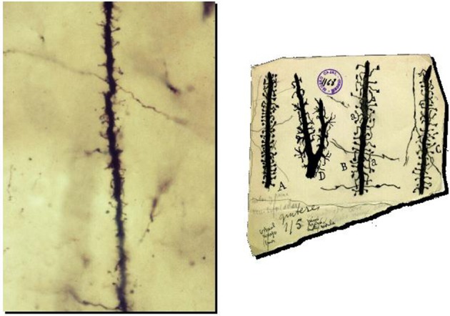
Figure 2.

Preparation and drawings of Cajal illustrating spines. Left: Photomicrograph of a dendrite of pyramidal neuron from one of Cajal's original preparations (Courtesy of Cajal Institute in Madrid). Right: Cajal drawings of spines from rabbit (A), 2 month old child (B), one month old cat (C) and cat spinal motoneuron (D). Reproduced with permission from “Herederos de Santiago Ramón y Cajal.”
