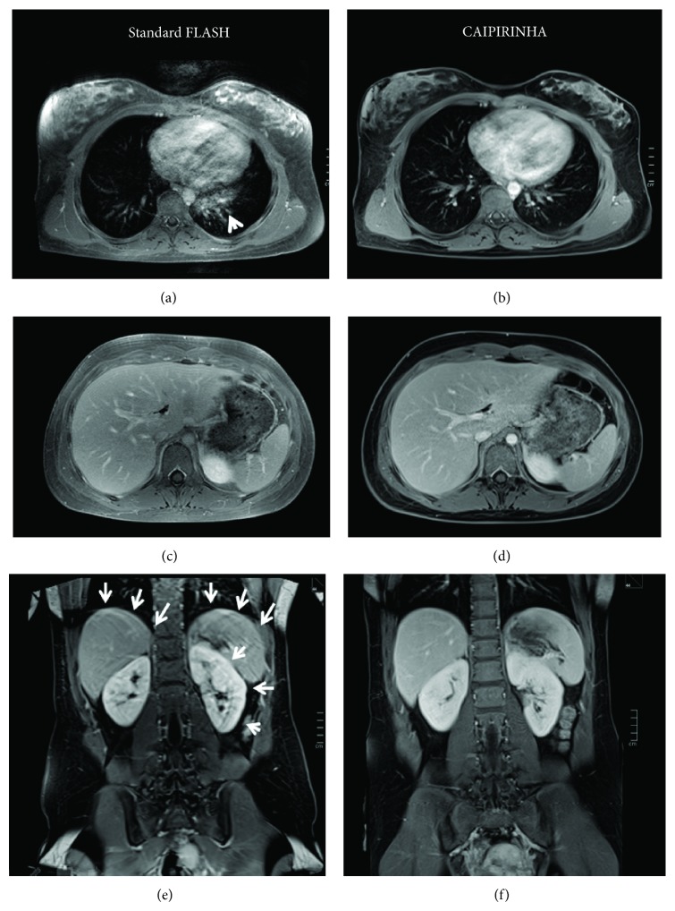
Figure 1.
Thoracic and abdominal MRI study in a 17-year-old female patient with Hodgkin's disease in complete remission. Images are displayed for contrast-enhanced fat-saturated standard FLASH (a, c, e) and CAIPIRINHA-accelerated T1w 3D FLASH (b, d, f) from the transverse thoracic image stack (a, b), the transverse abdominal image stack (c, d), and the coronal abdominal image stack (e, f). The sequences were scanned in turns on the respective level as standard FLASH first, followed by CAIPIRINHA. With CAIPIRINHA, there are markedly less retrocardial artefacts ((b), as compared to standard FLASH (a), arrowhead). While the transverse thoracoabdominal scans otherwise show excellent image quality with both FLASH and CAIPIRINHA, the patient could eventually no longer fully comply with the frequent breath-holds, so that the coronal FLASH image (e, arrows) is degraded by respiratory motion artefacts and shows blurring of contours along the diaphragm and the kidneys. The final acquisition of the coronal CAIPIRINHA sequence (f) again is virtually free of artefacts. The transverse abdominal scans were included in the study analysis and were consistently graded as IQ = 5 by both readers.

