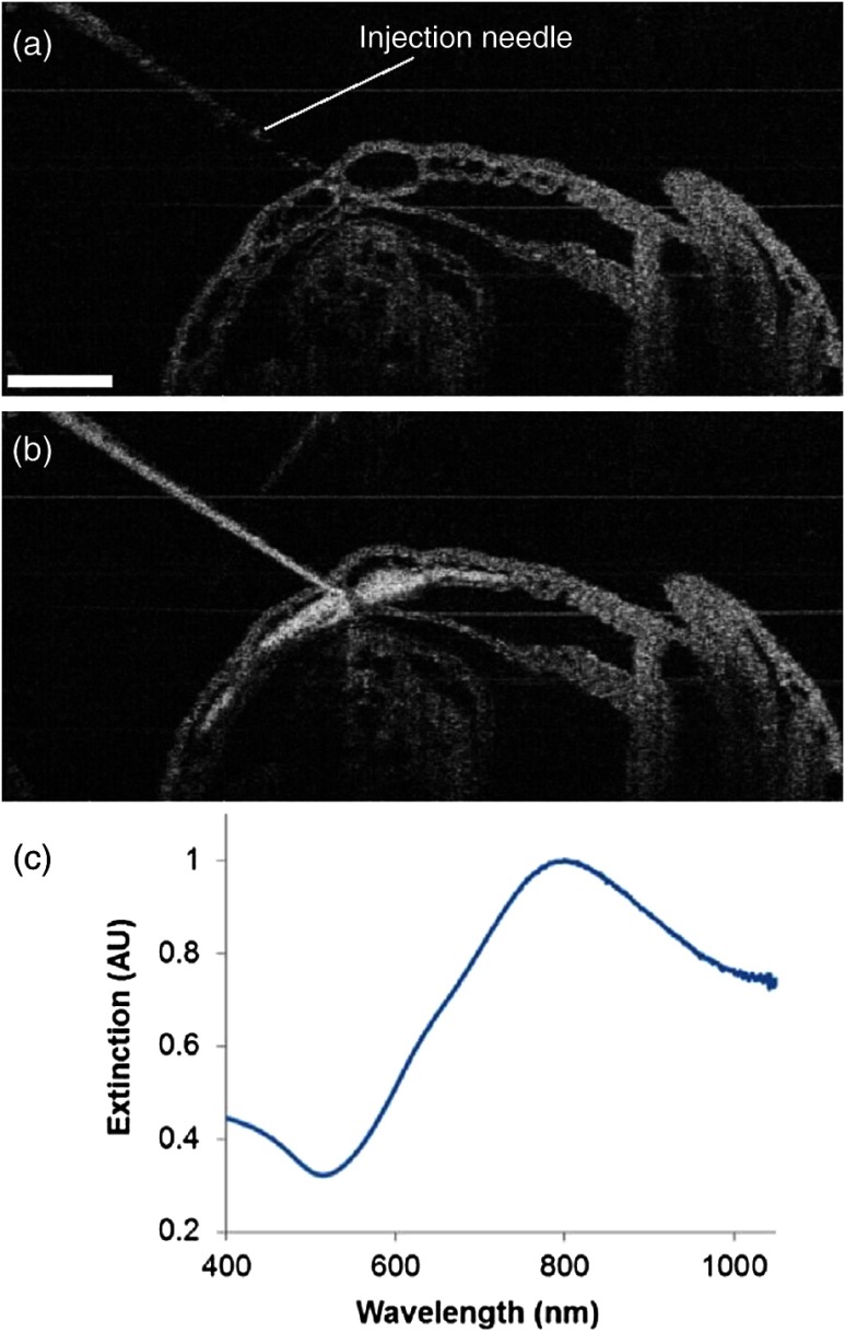
Fig. 4.
OCT-guided microinjection of gold–silica nanoshells in the yolk sac vasculature of a cultured live mouse embryo at early E8.5, when the heart just starts to beat. (a) Precise positioning of the injection needle into a vessel of the yolk sac that is only filled with plasma. (b) Microinjection of gold–silica nanoshells into the yolk sac vasculature as a contrast agent for the potential dynamic study of the cardiovascular formation and function before the onset of blood cell circulation. (c) Normalized extinction spectrum of the nanoshells used in this study showing the peak around 800 nm. The scale bar corresponds to and also applies for (b).

