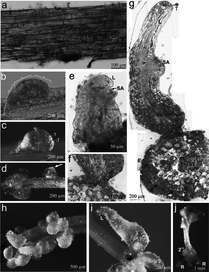
Fig. 3.
Light microscopy evidence for somatic embryo origin and development in Cyathea delgadii. a Numerous pro-embryos following several anticlinal, periclinal and inclined cell divisions of single epidermal cells of stipe explant cultured on hormone-free medium, in darkness. b Four-segmented somatic pro-embryo developed directly on the surface of stipe. c Trichomes located on one side of immature, multicellular somatic embryo. d Differentiation of the embryonic leaf (arrow). e A somatic embryo showing the first leaf and primordium of the second leaf (squashed specimen). f Junction between somatic embryo and initial stipe explant showing epidermal origin of embryo (semi-thin section stained with toluidine blue). g Longitudinal section of well-developed somatic embryo showing the first leaf, shoot apex and primordium of the second leaf, as well as transverse section of the stipe explant; 6 weeks of culture (arrows indicate amyloplasts). h Numerous somatic embryos with first leaf after 6 weeks growth. i Partly green, differentiated lamina of the first leaf of juvenile sporophyte. j Somatic embryo-derived young sporophyte showing extended lamina of primary frond and primordium of second leaf, as well as two roots, following development in the presence of light. C cortex, Ep epidermal cells, L first leaf, R root, SA shoot apex, T trichomes, Vc axial cylinder (color figure online)

