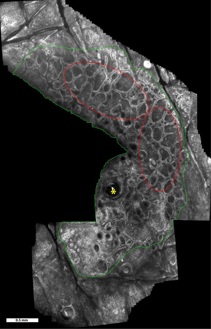
Figure 1.

Video mosaic of a benign melanocytic nevus over a 9.4 mm2 area, acquired in 50 seconds (~400 frames). Imaging was in the lower epidermis and at the dermo-epidermal junction. Features seen include normal skin (outside green boundary), nevus region (inside green boundary), hair follicle (yellow asterisks), and rings of dermal papillae (examples seen in the red ellipses).
