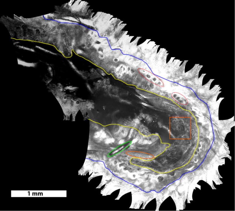
Figure 2.

Video mosaic of residual BCC margins, obtained intra-operatively in the surgical wound on a Mohs patient, over an area of 15.8 mm2, acquired in 40 seconds (~400 frames). Structures seen include dark nuclei within the honeycomb pattern of keratinocytes in the surrounding intact epidermis (outside blue boundary, appears saturated here), exposed epidermis in the wound (between blue and yellow boundaries), in which the nuclei (examples in orange oval) appear bright due to the application of aluminum chloride for contrast enhancement13, and the underlying papillary and reticular dermis (inside the yellow boundary). Other structures, such as collagen bundles in dermis (for example, inside orange square), rings of dermal papillae (for example, red ellipses) and hair shaft (green ellipse) can be seen in the mosaic.
