
FIG. 2.
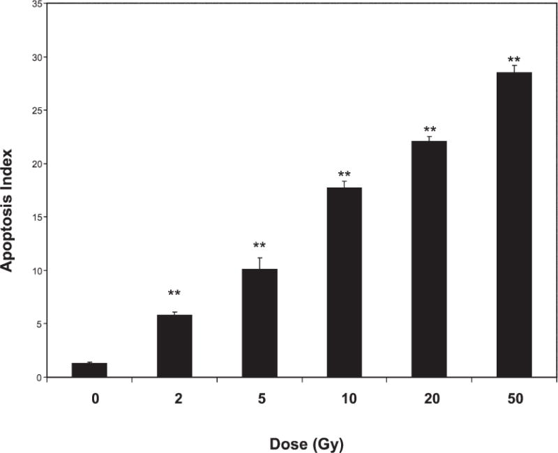
Apoptosis of endothelial cells after X irradiation. Cells (passages 5–8) were sham-treated (0 Gy) or exposed to various doses of X rays; 6 h later, the number of cells undergoing apoptosis was determined. Differences between sham-treated and irradiated cells were compared using Student’s t test (**P < 0.001). Shown are pooled data from treatments performed in duplicate per experiment in three separate experiments. Error bars are SE.