
Figure 9.
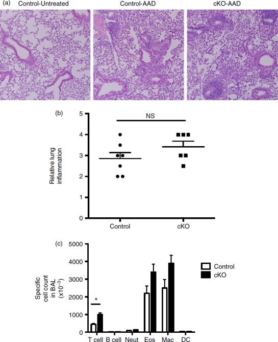
Inflammatory responses in regulatory T (Treg) cell-specific conditional Bcl6−/− mice after induction of allergic airway disease. Treg-specific conditional Bcl6−/− (Bcl6Foxp3−/−) (conditional knockout; cKO) mice and control Bcl6+/+ (Bcl6Foxp3+/+) mice were induced to develop allergic airway inflammation. Mice were primed with ovalbumin (OVA) antigen in Alum adjuvant intraperitoneally, and then challenged with OVA intranasally for 5 consecutive days, as described.21 (a) Representative lung histology on haematoxylin & eosin stained sections showing airway inflammation after disease induction. Control-Untreated is a control Bcl6+/+ mouse without any disease induction. Control-allergic airway disease (AAD) and cKO-AAD are control and cKO mice, respectively, induced to develop allergic airway disease. Strong infiltrates of inflammatory cells are observed around the airways of the AAD mice (b) Lung inflammation scores for Control-AAD and cKO-AAD mice, where the severity of the inflammatory infiltrate was analysed as described in Materials and methods. Each point shows one mouse. (c) Quantification of brochoalveolar lavage (BAL) cells obtained from Control-AAD and cKO-AAD mice. BAL cells were counted and assessed for specific cell types by flow cytometry. Graph shows average number of specific BAL cell types in Control-AAD and cKO-AAD mice. Neut, neutrophils; Eos, eosinophils; Mac, macrophage; DC, dendritic cells. n = 6 or n = 7. The experiment was performed twice with similar results obtained each time. NS, not significant (P > 0·05), *P < 0·05.