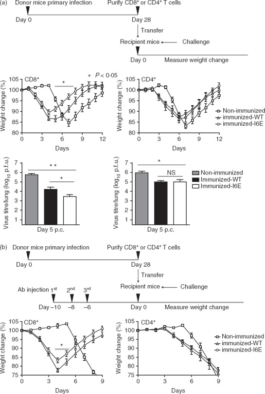
Figure 10.

CD8+ T cells from vN1.I6E-infected mice confer enhanced protection. (a) Mice were infected intradermally with vN1.WT or vN1.I6E or mock infected and either CD8+ or CD4+ cells were isolated 28 days post-infection and 106 cells were transferred into naive recipient mice. The recipient mice were challenged 6 hr later with 3 × 10³ plaque-forming units. of vaccinia virus (VACV) WR and weight change (middle panels) was monitored. *P < 0·05, n = 5. Lower panels show virus titres in the lungs 5 days post-challenge. Data are mean titre ± SEM, *P < 0·05, **P < 0·01. NS = non-significant. (b) As (a), except the naive recipient mice were depleted for CD8+ or CD4+ T cells by administration of monoclonal antibody at 10, 8 and 6 days before transfer of cells. CD4+ T cells were transferred to mice depleted of CD4+ T cells, or CD8+ T cells were transferred to mice depleted of CD8+ T cells. *P < 0·05, n = 5. Results are expressed as mean ± SEM.
