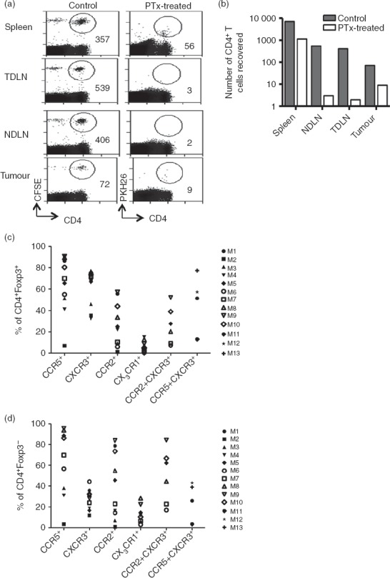
Figure 6.

Intra-tumoural recruitment of CD4+ T cells is chemokine dependent. Mice received 4 × 10⁷ splenic CD4+ T cells isolated from spleens of tumour-free Thy1.1 mice, half of which were treated with 25 nm Pertussis toxin and then labelled with PKH26 (desensitized fraction). The other half were left untreated (control) and labelled with CFSE. After 24 hr, spleen, non-draining lymph nodes (NDLN), tumour-draining lymph nodes (TDLN) and tumour were harvested, stained and analysed by flow cytometry. (a) Plots showing numbers of cells recovered from a representative mouse. (b) Absolute numbers of adoptively transferred cells that were recovered from each organ of a representative mouse. This experiment was carried out on two separate occasions with similar results. (c, d) Single cell suspensions prepared from tumours, spleens and lymph nodes were stained for various chemokine receptors and analysed by flow cytometry. Proportions of tumour-infiltrating regulatory T cells (c) or conventional T cells (d) expressing various chemokine receptors are shown as a percentage of CD4+ T cells. Each symbol represents a tumour from an individual mouse, M1–M13.
