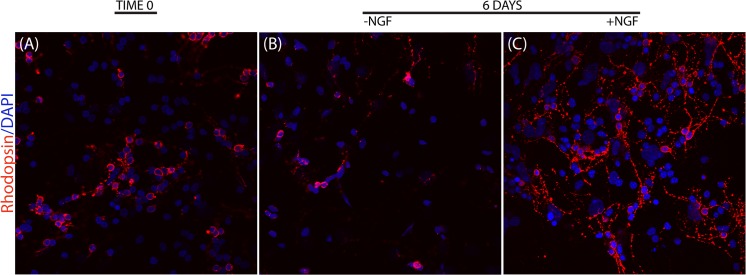
Fig 2. NGF effect on primary cultures of retinal cells.
Retinas were dissected out, dissociated and plated according to a standard procedure. Representative confocal images of p10 retinal cells expressing Rhodopsin at baseline (A) or after 6 days of culturing with medium alone (B) or supplemented with 50ng/ml NGF (C). Individual cells were attached as early as 12 hrs from seeding (A). NGF exposed cultures (C) retained retinal cells number, with respect to initial cultures (A) and showed an higher cell number with respect to in untreated ones (B). The red staining is Rhodopsin immunoreactivity in photoreceptors having blue nuclear counterstaining (DAPI). Magnification: x200

