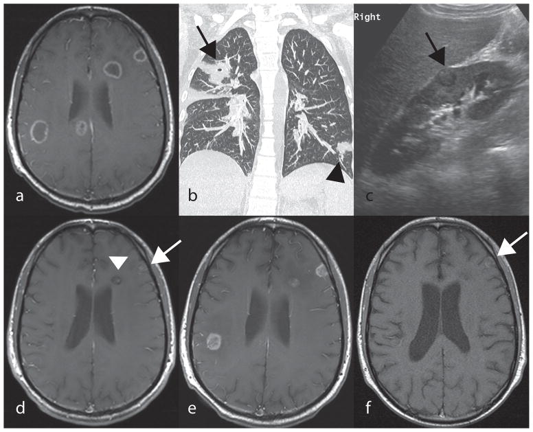
Figure 1.
A,D,E,F: Post contrast axial T1 spin echo of the brain. B: Coronal CT of the chest. C: Ultrasound of the right kidney (long axis). A: Multiple ring enhancing lesions within the periventricular white matter and peripheral cortex of the brain seen at presentation. B: Nodular lung opacity within the left lower lobe (black arrowhead) and a cavitating lung opacity in the right upper lobe (black arrow). C: Round peripheral renal lesion (black arrow) with a hypoechoic rim. D: Significant decrease in number and size of brain lesions (white arrow and arrowhead) after 22 months of antifungal treatment E: Relapse with increase in size of brain lesions showing now rim enhancement following one month off voriconazole therapy due to chemotherapy. F: Significant decrease in number and size of brain lesions showing intrinsic T1 hyperintensity in one of the lesions (white arrow) with absence of enhancement prior to successful HCT transplant, following four months of therapy from relapse in Figure 3E, above.

