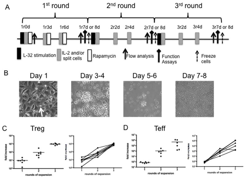
Fig. 3. Protocol for ex vivo-expansion of cynomolgus Treg using artificial APC and the assays performed.

(A) Treg expansion protocol. Freshly-sorted Treg (CD4+CD25hiCD127−/lo) or conventional Teff (CD4+CD25−CD127hi) were stimulated with anti-CD3-loaded artificial APC (L-32 cells) in the presence of 300 U/ml recombinant human IL-2 for 7–8 days. Rapamycin (100 ng/ml) was also added to the cultures for the first round. The cells were harvested and expanded for an additional 2 rounds, as in the first round, except that no rapamycin was added. At the end of the 2nd round (2r7 or 8 days), a portion of the cells was frozen for future expansion. r: round of expansion; d: days in a round (B) Appearance of the cultures during the course of round 1. Treg (arrow) appear to attach to L-32 cells on day 1. Cultured cells were transferred to larger vessels after day 3–4. Homogeneous appearance of expanded Treg on days 7–8, with minimal/no L-32 cell contamination. (C, D) Fold expansion of Treg and Teff at the end of each of 3 rounds (results of 6 experiments).
