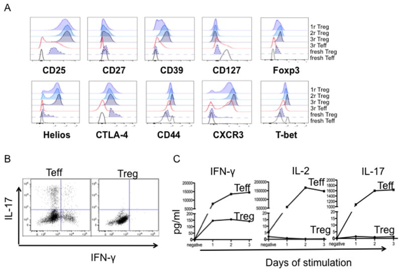
Fig. 4. Phenotype and function of expanded cynomolgus Treg and Teff from each round.

(A) Expanded Treg and Teff were stained for Foxp3, CD25, CD27, CD39, CD44, Helios, CTLA-4, CD127, CXCR3 and T-bet at the end of rounds 1, 2 and 3 of expansion. PBMC were used as controls: fresh Teff (CD4+CD25−CD127+); fresh Treg (CD4+CD25+CD127−). Data are representative of 3 experiments. (B, C) Expanded (3 rounds) Treg and Teff were stimulated with L-32 cells for 3 days. (B) On day 1, some cells were further activated for 4h with PMA and ionomycin in the presence of GolgiStop (BD Bioscience), followed by staining with LIVE/DEAD fixable dye (Molecular Probes: Invitrogen) and fluorescent-tagged Ab against CD4. Intracellular IFN-γ and IL-17 expression were detected according to the eBioscience Intracellular Foxp3 Staining Protocol. (C) Supernatants on days 1, 2, and 3 were collected and IFN-γ, IL-2 and IL-17 levels quantified. Data are representative of 2 experiments.
