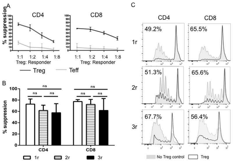
Fig. 5. Expanded cynomolgus Treg strongly suppress CD4+ and CD8+ T cell proliferation.

(A) Cynomolgus Treg expanded for 2 rounds were tested for their suppressive activity in CFSE proliferation assays using various responders (CD2+ T cells): Treg ratios, as described in the Materials and Methods. Percent Divided were calculated using FlowJo software. Treg function was expressed as percent suppression of autologous CD4+ or CD8+ T cell proliferation calculated using the formula: (percent divided T cells without addition of Treg or Teff – percent divided T cells with Treg or Teff)/percent divided T cells without addition of Treg or Teff X 100%. Expanded Treg (black lines) showed strong suppressive activity when added to bead-stimulated T cells, whereas expanded Teff (gray lines) did not. Data are representative of 2 independent experiments. (B, C) Cynomolgus Treg expanded for 1, 2 or 3 rounds were tested for their suppressive activity in CFSE proliferation assays using responder (PBMC): Treg ratio of 2:1. (B) Percent suppression from 4 independent experiments was plotted as bar graph. (C) Representative histogram overlay of proliferation with (black line) and without (grey shading) addition of Treg. All three types of Treg showed comparable suppressive capacity when added to bead-stimulated PBMCs.
