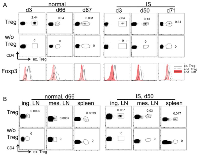
Fig. 7. Persistence of ex-vivo expanded Treg in peripheral blood, LN and spleen after systemic infusion.

Autologous Treg expanded from frozen 2nd round Treg were labeled with CFSE or VPD450 dye and infused into normal or immunosuppressed (IS) monkeys. On the indicated days post infusion, 1ml blood (A, n=2) and/or lymphoid tissues (B, n=1) were assessed for the presence of infused cells (ex. Treg) by flow cytometry. Samples from monkeys without receiving cells labeled with the same dye were used as negative controls. Blood samples were further tested for Foxp3 expression in infused cells (CD3+CD4+CD25+CD127−Dye+, black solid line), endogenous (end.) Treg (CD3+CD4+CD25+CD127− Dye−, black dashed line) and end. Teff (CD3+CD4+CD25−CD127+, shaded profiles). Histogram overlay of the three populations is shown (A).
