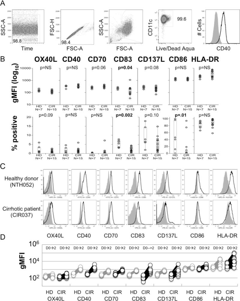
Fig. 3.

Baseline and post-maturation phenotyping of monocytes from cirrhotic and healthy donors. (A) Gating strategy used to phenotype MoDCs. (B) Comparison of the geometric MFI and frequency of positive expression of OX40L, CD40, CD70, CD83, CD137L, CD86 and HLA-DR on CD14+ monocytes prior to maturation in cirrhotic patients (CIR) and healthy donors (HD). p-Values determined by Wilcoxon Rank-Sum test. (C) Representative histograms showing expression of OX40L, CD40, CD70, CD83, CD137L, CD86 and HLA-DR from cirrhotic subject and healthy donor pre-stimulation (grey shaded) and post-maturation (black line). (D) Pre- and post-maturation changes of geometric MFI of OX40L, CD40, CD70, CD83, CD137L, CD86 and HLA-DR costimulation markers on monocytes and MoDC. No significant differences were identified between CIR and HD.
